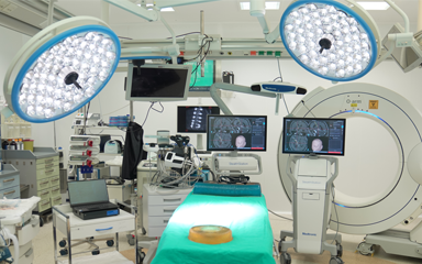
Hybrid Operating Room

At Liv Hospital Brain and Nerve Surgery Clinic, hybrid operating rooms are used for many disease groups and trauma surgery in brain, spinal cord and spine surgery. Neurovascular surgery of vascular diseases in the brain or spinal cord such as aneurysm (bubble), arteriovenous malformation (AVM), brain tumor or functional stereotactic surgery and spinal implantation surgery are some of these.
O-ARM CT (0-ARM TOMOGRAPHY)
Computerized tomography can be taken during surgery with the mobile tomography device, which is a new generation imaging system. With this technology, 2- or 3-dimensional imaging can be performed at the time of surgery in both brain and spine-spinal surgery. Moreover, in three dimensions at a 360-degree angle, in as little as 13 seconds. The system also has a robotic positioning feature. In this way, the device can be approached to the patient table very quickly in the operating room and a tomography can be taken. Since it takes images in one go, images with the same image quality can be viewed throughout the surgery with one-third less radiation than other standard tomography devices.
Areas where it is most used:
- Spinal deformities due to aging
- Compression in the spinal canal
- In spinal tumors
- Spinal curvatures in childhood and adolescence
- In some developmental diseases of the spine
- Screwing (platinum placement) in fractures and dislocations due to trauma
While the success rate of spinal screwing surgeries performed with O-Arm technology, which can take 3-dimensional tomography images, increases, the margin of error is also zero. In addition, the chance of surgery success can be further increased by using the neuronavigation system, which works in harmony with the O-Arm CT device and can show all targets with high precision, at the same time.
With a precision of 1-2 mm, the risk of injury to the spinal cord and nerve roots is almost eliminated and a safer surgery is performed. Thanks to the O-Arm CT system, tomography is performed under sterile conditions at the last stage of the surgery, minimizing undesirable situations that require the patient to undergo re-operation. The patient stands up in a short time and is discharged quickly.
Advantages of spinal screw surgery performed with the O-Arm device:
- It provides critical information to the surgeon at every stage, and the risk of repeat surgery is reduced.
- The patient receives less radiation.
- With a smaller surgical incision, it provides the patient with a faster recovery and reduces bleeding.
- The system minimizes the major risks of complex surgeries.
- It reduces the risk of infection. The risk of screw-related paralysis is reduced to the lowest level.
NEURO-NAVIGATION SYSTEM
It calculates the coordinates of a target to be visited in brain or spinal cord surgery with a system similar to GPS technology and allows the surgeon to see 3-dimensional scanned images. With "neuronavigation", the targeted lesion in neurosurgery is approached with a high degree of accuracy (precision less than 1 mm). This minimizes the damage that may occur to healthy tissue. Before the surgery, the patient's MRI and/or CT scan is taken and transferred to the navigation device. Thus, surgery is planned by seeing various risk areas in the patient's brain and spine with real-time navigation.
In all cases of small and deep-seated tumors and similar cases, stereotaxic biopsy can be performed using only a small entry hole. In other words, a thin needle is directly reached to that area millimetrically and a tissue sample is taken. And of course, as a result of the pathological examination of this tissue sample, it is determined which treatment method the patient will be treated with.
Advantages:
- It moves flawlessly over delicate anatomy and avoids critical structures.
- The surgery is performed within more minimally invasive limits (surgery time is shortened with a small surgical incision).
- It provides safer surgery by not disrupting the patient's healthy anatomy.
NEW GENERATION FLUORESCENT FILTER MICROSCOPE
With new generation microscopes and special light filters, tumor tissue stained with fluorescence can be easily distinguished from normal nerve tissue. Therefore, thanks to the special operating microscope combined with neuronavigation, maximum tumor removal is achieved with a smaller incision and with the least error. Apart from tumor surgeries, this system is also used in surgeries of cerebrovascular diseases.
For example, thanks to the special substance given intravenously to the patient during surgery, the filter of the microscope can be adjusted and the vessels of the brain can be seen, that is, cerebral vascular angiography can be performed. In this way, the vascular network of the brain is monitored before the surgery is completed. This increases the chance of surgical treatment of cerebrovascular disorders more safely.
