PET - CT
PET-CT is a system that combines PET (positron emission tomography) and CT (computed tomography) to evaluate functional images with cross-sectional anatomical information.
What is PET-CT?
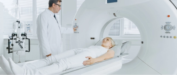
PET-CT is a system that combines PET (positron emission tomography) and CT (computed tomography) to evaluate functional images with cross-sectional anatomical information. While PET provides information about the function and metabolism of cells, CT provides anatomical data such as size, location and density.
In oncology patients, it is used to differentiate between benign and malignant masses, to stage cancer, to determine the degree of malignancy of the tumour, to determine the spread of the tumour within the organ, to determine whole body metastases, to detect recurrence, to select the treatment method, to evaluate the response to treatment, to determine the actual tumour volume for radiotherapy planning and to ensure that the right place is irradiated with the right dose. All evaluations are supported by numerical information. In oncology, PET-CT is a method that provides more information than the sum of all other imaging methods by imaging the whole body at once.
PET-CT at Liv Hospital
PET-CT at Liv Hospital is an advanced imaging technology that combines positron emission tomography and computed tomography. This technology helps to diagnose and plan treatment by providing a detailed view of cellular activity and changes in anatomical structures within the body. PET-CT is used in many medical fields such as oncology, cardiology and neurology, as well as for cancer screening and evaluating treatment response, the determination of the actual tumor volume in the planning of the radiotherapy used. Supports all assessments with numeric information. In the oncological patient group, PET-CT is a method that displays the whole body at a time and provides more information than all other imaging methods.
Usage Areas
Nuclear medicine applications are mostly used in the oncological sense to scan the whole body. To determine the main focus in cancer-diagnosed patients, to determine the main focus in patients diagnosed with cancer, to determine the prevalence of the nodules or masses previously diagnosed in patients, to determine the prevalence of other body regions. In the evaluation of the response, recurrence (renewal) or suspicion of metastasis (spreading elsewhere), recently in the planning of radiotherapy and to be irradiated live cancer tissue more accurately determine and contribute to the protection of healthy tissues.
It is used in the differential diagnosis of solitary pulmonary nodules, brain tumors, pancreatic masses and adrenal gland masses.
In the evaluation of treatment response; In the event of a successful response, chemotherapy may be decided earlier to continue chemotherapy or, if necessary, to replace chemotherapy drugs (for example, if the cancerous tissue resists treatment).
It has an important role in the investigation of the presence of live muscle tissue before the intervention of the stent, such as by-pass and stent, in the selected heart patients and thus to determine whether these interventions are beneficial.
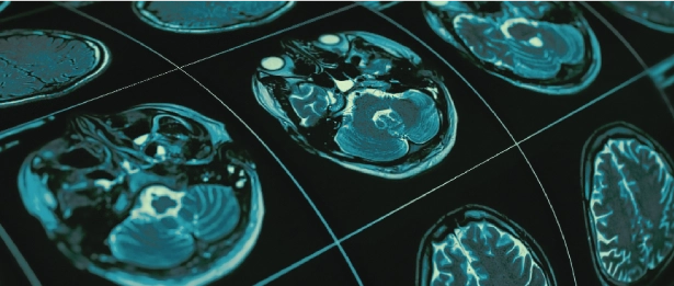
It is used in psychiatry and neurological sciences to show brain glucose metabolism, oxygen consumption and blood flow with different agents, to detect epilepsy foci, and to monitor many nervous system related receptors and molecules. It can now be used to evaluate the status of dopamine receptors in Parkinson's patients and to monitor response to treatment. It can also be applied in the differential diagnosis of other movement disorders.
What are the preliminary preparations for PET-CT patients?
For PET-CT, patients should be hungry for 6 hours before the examination. Patients may take oral antidiabetic or non-insulin drugs. In diabetic patients, image quality may be poor because of the competition of blood glucose with FDG. For this reason, blood glucose level is determined before FDG injection and it is generally not recommended to perform blood glucose levels above 180 mg / dL.
How is PET-CT imaging performed?
During PET-CT imaging, the resting patient is injected intravenously with a special radiopharmaceutical called FDG (Fluorodeoxyglucose). After waiting for approximately 60 minutes for FDG to accumulate in cancerous tissues, an approximate 20-25 min. If necessary, a second imaging can be performed after 2 hours.


.webp)

.webp)
