


What is Breast Biopsy?
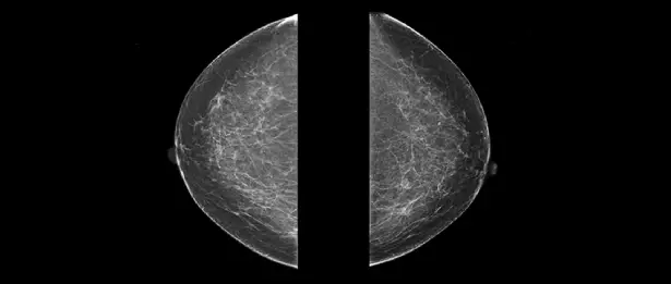
Imaging studies such as mammography, ultrasound and MRI (magnetic resonance imaging), which are frequently performed along with breast examination, may cause the doctor to suspect that the person may have breast cancer. However, the only way to know for sure is to take a sample of the tissue in the suspicious area and examine it under a microscope. A biopsy is a minor procedure to remove tissue from a relevant area of the body. If your doctor feels something suspicious in your breast or sees something suspicious on an imaging study, he or she may order a biopsy. The tissue sample is examined by an expert pathologist for the presence of cancer cells. If cancer is present, then the pathologist evaluates the characteristics of the cancer. The pathologist's findings are prepared in the form of a pathology report.
Breast Biopsy at Liv Hospital
Liv Hospital is a healthcare institution that successfully performs diagnostic procedures such as breast biopsy. Breast biopsy is a procedure that allows abnormal cell or tissue samples detected in the breast to be taken and examined in a laboratory environment. This method is an important step in diagnosing breast cancer and identifying other breast diseases. Liv Hospital offers patients a safe and effective biopsy experience with its experienced specialists and modern medical equipment.
How Many Types of Biopsies Are There?
There are various techniques for performing a biopsy, and your doctor will likely recommend the simplest and easiest method possible for you (fewest incisions, minimal scars). However, the choice of procedure depends on your current situation. Biopsy is basically in two ways; It can be done as a small surgical procedure in which a piece of tissue is removed by inserting a needle into the breast skin or by incising the breast skin and removing some or all of the suspicious tissue.
Fine Needle Aspiration Biopsy (FNAB): It is the biopsy method that requires the least amount of intervention into the body and usually leaves no scars. First, local anesthesia is given and numbed to the area where the needle will be inserted into the breast, and then the interventional radiologist or surgeon uses a fine needle with a syringe to take cell samples from the suspicious area.
In cases where the mass is felt comfortably, there may be no need for any auxiliary imaging method. However, in cases where the mass cannot be felt or if there is an area of the mass that specifically requires sampling, the interventional radiologist or surgeon can use auxiliary imaging devices to direct the needle to the right place. If ultrasound is used for this purpose, it is called "ultrasound-guided biopsy" or if mammography is used, it is called "stereotactic biopsy". In ultrasound-guided biopsy, the doctor directly watches the needle tip enter the relevant area in the ultrasound image. In stereotactic biopsy, the location of the mass is calculated by taking mammography images from various angles and the doctor takes cell samples by inserting a syringe needle into the calculated location.
With fine needle biopsy, a cell sample is taken, not a tissue sample. In this method, although it is possible to see cancerous cells, tissue identification (milk bladder or milk duct) cannot be made.
Thick Needle (Tru-cut) Biopsy (CRB): It is performed with a needle with a thicker diameter and automatic pulsation than fine needle aspiration. After the area where the needle will be inserted into the breast is local anesthetized and anesthetized, the interventional radiologist or surgeon takes several cylindrical tissue samples from the suspicious area. The tissue collection process is repeated 3-6 times to obtain sufficient samples. Usually there is no scar left on the skin.
In cases where the mass cannot be felt or if there is an area of the mass that specifically requires sampling, the interventional radiologist or surgeon can use auxiliary imaging devices to direct the needle to the right place. In cases where the tissue is proven to be cancerous and additional surgery is required, a small metal clip may be placed on the breast to mark the biopsy site. This clip is left inside the breast and has no harm to the body. If surgery is decided after the biopsy, the clip is removed during the procedure.
In addition to providing rapid results without significant discomfort or scarring, both fine and thick needle biopsies provide you with the opportunity to discuss treatment options with your doctor before any surgery. However, the risk of “false negative” results is higher in needle biopsy. The result is that there is no cancer when there is actually cancer. This is probably because a smaller sample of tissue is taken with a needle than with a surgical biopsy, and cancer cells may not be removed. Your doctor may recommend an open biopsy instead of a needle biopsy or during follow-up. You can decide together what is best for you.
Vacuum-Assisted Breast Biopsy: It is a newer method of doing a breast biopsy. Unlike a thick needle biopsy, a vacuum-assisted biopsy involves a special needle that is inserted through the skin only once. Additionally, more tissue may be removed than is removed in a needle biopsy.
For a vacuum-assisted breast biopsy, you lie face down on a special examination table that has slots into which you can place your breasts. First, a local anesthetic injection is given to numb the breast. Under the guidance of mammography (stereotactic-guided biopsy) or ultrasound, the surgeon or interventional radiologist inserts a biopsy probe into the suspicious area of the breast. The vacuum then begins to draw the tissue into the catheter. A rotary cutting tool removes tissue samples and then moves them through the catheter to a collection area. The surgeon or interventional radiologist can then rotate the probe around its axis to obtain additional tissue samples from the suspicious area. This process can be repeated 8-10 times until the entire area of interest is completely sampled.
In some cases, a small metal clip is placed to mark the area if a repeat biopsy is needed in the future. The clip is placed inside the breast and does not cause any pain or harm. If a surgical procedure is planned after the biopsy, the clip is removed during this procedure.
Open Sectional Biopsy (Incisional Biopsy): Open cross-sectional biopsy is more like normal surgery. After the breast is anesthetized with local anesthesia, the skin is cut with a scalpel and a piece of tissue is removed and sent for examination. In this biopsy, only a part of the suspicious mass or area is removed, not the entire area.
If the mass or suspicious area cannot be felt, as in a needle biopsy, there may be a need to use mammography or ultrasound to find the correct spot. It can be used in a process called wire marking. A small slotted needle is inserted through the breast skin into the suspicious area under mammography or ultrasound guidance. A thin wire is passed through the needle and placed in the relevant area. The needle is then withdrawn and the wire remains in place. This way, the surgeon can find the right spot with wire guidance during the biopsy.
If the results of a needle biopsy are inconclusive, meaning that the results are unclear or inconclusive, or if the suspicious area is too large to be easily biopsied with a needle, your doctor may recommend an open cross-sectional biopsy. As with needle biopsy, there is some possibility of false negative results (cancer but no cancer) with open section biopsy. However, the results are obtained quite quickly. Since it is a surgical procedure, open cross-sectional biopsy is an approach that requires a skin incision, leaves a scar, and requires a longer recovery time compared to needle biopsy.
Open Holistic Biopsy (Excisional Biopsy): Open holistic biopsy is a surgical procedure performed to remove the entire suspicious area or mass in the breast. In addition to removing the suspected cancer, some normal breast tissue is usually removed around the mass or suspicious area.
If the mass or suspicious area cannot be felt, as in an open cross-sectional biopsy, there may be a need to use mammography or ultrasound to find the correct spot. Wire marking can also be used to mark the correct area to be biopsied.
Open holistic biopsy is the most reliable way to obtain a definitive diagnosis without a false negative result. Also, having the entire mass removed may give you some peace of mind. However, open holistic biopsy is similar to open cross-sectional biopsy. It is an approach that leaves scars and requires a longer recovery time. It can be done with local or general anesthesia.
What Should Be Considered Before Biopsy?
Biopsies are not a medical emergency and an appointment can be made at a time convenient for you. However, most patients want to have their biopsy done immediately to feel relieved.
Before having a biopsy, discuss the following with your doctor:
• Review the results of mammography or other imaging tests performed on you.
• Ask him to show you the problem area.
• Ask what type of biopsy is recommended and why that type of biopsy is recommended.
• Discuss how and why a biopsy should be performed.
• Ask all the questions on your mind.
• Fill out the consent form that you must sign.
• Ask when and how you will receive your biopsy result.
Within a few days to a week, your doctor will give you your pathology report and explain what the result is.
What kind of treatment is applied at Liv Hospital?
Patients who apply to our hospital for breast cancer screening or diagnosis and who are considered for breast biopsy as a result of the necessary examinations and tests are recommended a biopsy by their doctors, the reason for the biopsy and its options are discussed and a decision is made together. Then, if a needle biopsy is decided, the patient is referred to the Interventional Radiology department for a needle biopsy appointment and procedure. After the biopsy process is completed in the Interventional Radiology department, the sample taken is sent to the Pathology department for examination.
If an open surgical biopsy is to be performed, the patient and doctor decide together whether this procedure will be performed under local or general anesthesia. If it is decided to perform it with local anesthesia, the patient is given an appointment for the day of the procedure and the procedure is performed on the day of the procedure by the patient's doctor, a General Surgery Specialist experienced in breast diseases. If it is decided to perform it under general anesthesia, the patient is given an appointment for the day of the surgery and after the necessary preparations for the surgery are made, the procedure is performed on the day of the surgery by the patient's doctor, a General Surgeon who is experienced in breast diseases. The tissue samples taken are immediately sent to the Pathology Department for examination.
After the pathology result is known, the patient is called by his doctor and informed about the result.
Which Physicians and Clinics are Collaborated with?
Radiology and Pathology Specialists work in collaboration with General Surgery Specialists who specialize in breast diseases.
* Contents of this page is for informational purposes only. Please consult your doctor for diagnosis and treatment. The content of this page does not include information on medicinal health care at Liv Hospital

